Benjamin Auer, PhD
Director, Nuclear Medicine Physics
Instructor
Marie Foley Kijewski, Sc.D.
Associate Physicist
Associate Professor
The nuclear medicine physics group works on a variety of research projects, most of which are related to task-dependent optimization of systems for improved nuclear medicine imaging, including optimization of
- collimation,
- reconstruction algorithms, and
- methods of compensating for a variety of physical effects
In addition, we have been directly involved in supporting both clinical and preclinical research projects in collaboration with other physicians and scientists within the radiology department, in other departments, and with outside institutions.
We are actively engaged in teaching radiology residents and fellows in the cardiovascular imaging and nuclear medicine training programs. We have clinical service responsibilities within the radiology department and the nuclear medicine division, including instrument quality assurance and acceptance testing of new imaging and detector systems.
Selected Research Projects
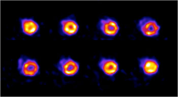
- Physical limits of quantitative ECT. (Kijewski MF; 5R01-EB000802). In this research project, funded by NIH for 18 years, we have most recently developed and evaluated a hybrid collimation system for brain SPECT consisting of an ultra-short cone-beam collimator on one gamma camera paired with a fan-beam collimator on the opposing camera. We have investigated the utility of time-of-flight PET imaging for improving quantitative task performance in cardiac and brain imaging.
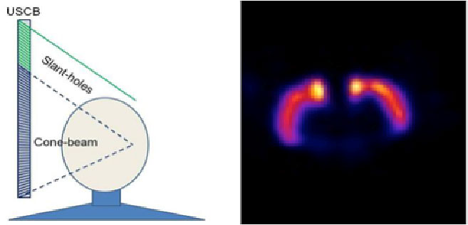
Left: Ultra-short cone-beam collimation concept. Right: Reconstruction of striatal phantom. |
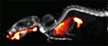
- Quantitative ECT for complex imaging tasks. (Moore SC; 5R01-EB001989) The major goals of this project, funded by NIH for 14 years, have been: to jointly optimize SPECT collimation and reconstruction; to develop a novel method of correcting for the partial-volume effect in SPECT and PET imaging; to implement and evaluate a simultaneous dual-radionuclide acquisition method for SPECT imaging of osteomyelitis; and, to develop a fast forward-projection algorithm for reconstruction of Y-90 bremsstrahlung photons.
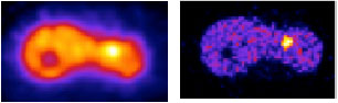
Left: Y-90 bremsstrahlung SPECT. Right: Y-90 PET image. |
- Preclinical cardiac imaging package for clinical SPECT systems. (Metzler SD and Moore SC; 1R01-HL111883-02). In this project, we are developing, in collaboration with researchers at the University of Pennsylvania, a very high-resolution, high-sensitivity system for cardiac SPECT imaging of mice. The approach is based on the use of a clinical triple-head SPECT camera in conjunction with two collimator tubes, each of which will contain many pinholes. One collimator tube is used for scout imaging of a mouse placed inside the tube, the other will be used for high-resolution imaging of a targeted organ of interest, e.g., heart, brain, etc.
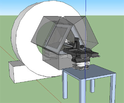
Perspective drawing of our new microSPECT scanner currently under development. |
- The nuclear medicine physics group provides support for the BICOR small-animal molecular imaging facility in Thorn 307-309, including the facility's microPET/CT and microSPECT scanners.
Small-animal PET and PET/CT images acquired in the BICOR small-animal imaging facility
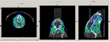
PET/CT images of glioma in a mouse using 18F-FLT. |
Gated FDG PET image of a rat heart. |
Small-animal SPECT images acquired in the BICOR molecular imaging facility
99mTc-MDP mouse bone scan (gray), registered to a 99mTc-sulfur colloid image (red). for evaluating mucociliary clearance of particles from the lungs. |
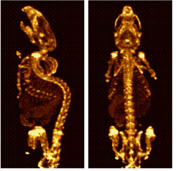
99mTc-MDP mouse bone scan |

