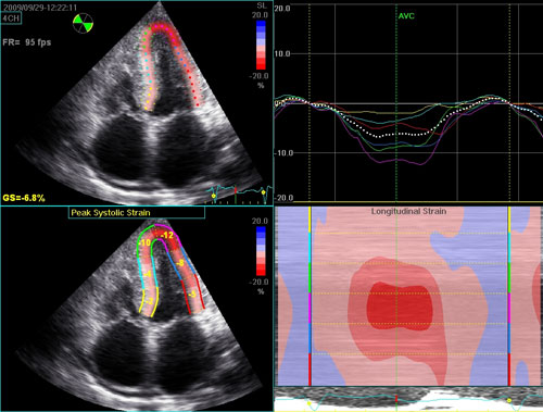
Typical echocardiogram for a patient with AL amyloidosis. There is biventricular wall thickening with normal cavity size, and bi-atrial enlargement. The atria are immobile, best noted on the apical 4-chamber view. Despite the normal left ventricular ejection fraction, longitudinal function is significantly impaired, as noted on the tissue Doppler and on the color map of longitudinal strain.

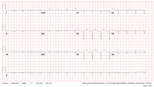
Electrocardiogram (Download PDF) from the patient illustrated in the video clips, showing classical features of cardiac amyloidosis. Note the low voltage (despite increased wall thickness on the echocardiogram) with left anterior fascicular block and an appearance suggestive of an old septal myocardial infarction (the patient had no epicardial coronary artery obstruction) with generalized poor R wave progression in the precordial leads.
For over a century, a leader in patient care, medical education and research, with expertise in virtually every specialty of medicine and surgery.
About BWH

