Image-guided percutaneous tumor ablation is a growing field world-wide. There are ongoing increases in the number of procedures and advances in both the technologies of imaging and the ablation devices. Our tumor ablation program uses a full array of several imaging techniques (ultrasound (US), computed tomography (CT), magnetic resonance imaging (MRI) and positron emission tomography (PET)) and therapeutic ablative modalities (alcohol, radiofrequency, cryoablation, microwave, irreversible electroporation). Our program is unique in this respect as compared against other centers that typically use one ablative modality and one or two imaging techniques; we use all of them.
Our interest and research efforts in image-guided tumor ablation have arisen from developments in the field of medical imaging that have made image-guided percutaneous interventions feasible. These developments include both improved image acquisition and advanced computer visualization of image data. In 1988, our group presented and published the first research on the use of MRI to visualize therapeutic thermal events in tissue. The initial focus was on MRI of experimental interstitial laser tissue coagulation and cryoablation. These were pioneering efforts of MRI image-guided thermal therapies and established a foot-hold in the history of the field.
The Tumor Ablation Program (TAP) was born out of the Department of Radiology’s Cross-Sectional Interventional Radiology (CSIR) service that officially commenced clinical operations in 1990 as a Departmental commitment to interventional radiology. The general interventional practice expanded steadily through the 1990’s with an increasing role in patient care, mirroring a nationwide trend.
The research efforts in MRI both fueled a growing interest in clinical applications and research of percutaneous thermal ablation of tumors. Technical developments and physical resources have always played vital roles for CSIR and TAP from the acquisition of a dedicated US system to the advent of CT fluoroscopy. However, a key development for TAP was the opening of the interventional MRI suite in 1994. The centerpiece of this suite was the first-of-its-kind, a “vertically open” interventional 0.5 Tesla scanner. After performing the first-ever fully MRI-guided needle biopsy, the principles and techniques of targeting and navigating with MRI were established and extended to percutaneous tumor ablations in 1996.
The first image-guided ablation performed in the open MRI scanner targeted a liver metastasis with interstitial laser thermal photocoagulation. In 1998, the range of treatment modalities was extended to include cryoablation for tumor ablation. The ablation service extended further with the addition of interstitial radiofrequency (RF) coagulation - done percutaneously under CT guidance. In 2000 and 2001, cryoablation and RF ablation (RFA) were applied outside the liver in the kidney under MRI and CT, respectively. Tumor ablation in the adrenal gland was added in 2001. The program’s first CT-guided RFA in the lung was first performed in 2001.
After more than five years of development, the program’s 100th ablation procedure was performed by the end of 2002; remarkably, the next 100 procedures were completed before the close of 2003! With new momentum, the program grew – over the years more milestones were reached, a weekly physician “Clinical Rounds” was implemented, as well as new clinic space identified for the ablation practice. In 2006, and since, cryoablation was performed under CT guidance. Also, a new wide bore CT scanner was installed for interventions providing more room for physician and patient alike. The 500th procedure was performed in mid-2007. We have performed our first ablation cases under PET/CT guidance, and have commenced to using microwave ablation - all in an effort to broaden our capabilities to address the challenges of our growing referral base of patients.
In 2011, BWH established The Advanced Multimodality Image Guided Operating (AMIGO) suite, a state-of-the-art interventional radiology and surgical research environment that houses a complete array of advanced imaging equipment, interventional, and surgical systems. Multidisciplinary teams of specialists use this equipment array and the unique design of the suite to efficiently and precisely guide treatment — before, during, and after surgery — without the patient or medical team ever leaving the operating room. This innovative operating and imaging research suite encourages collaboration among multidisciplinary teams of surgeons, interventional radiologists, imaging physicists, computer scientists, biomedical engineers, nurses, and technologists. Harnessing the benefits of advanced technology and an efficient three-room design, the AMIGO teams aim to develop and deliver the safest and most-effective state-of-the-art therapies in a patient-friendly environment.
Now with the AMIGO suite open as a new location for more procedures, the Tumor Ablation Program currently treats 6-8 patients each week in AMIGO. Our 3000th ablation procedure was performed in March, 2022.
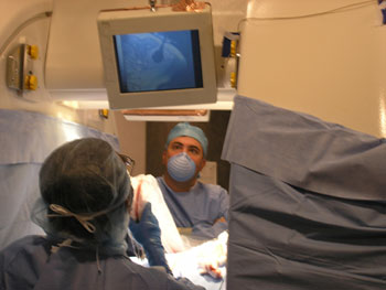 Kemal Tuncali, MD, views the iceball achieved during a percutaneous MRI-guided cryoablation in the original interventional MRI scanner. |
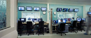 The interventional/surgical control room in AMIGO. |
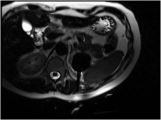 The Tumor Ablation Program has performed over 400 MRI-guided renal cryoablations. |
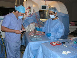 Preparing for a MRI-guided cryoablation in one of the first interventional MRI scanners ever built. |
| 1990 | CSIR opens: US, CT-guided interventions offered to patients |
| 1994 | Interventional MRI suite opens |
| First percutaneous MRI-guided procedure performed: MRI-guided abdominal mass biopsy (the first MR-guided biopsy in which entire procedure was performed under MRI-guidance) | |
| 1995 | CSIR patient volume increases steadily through the 1990s |
| 1996 | First percutaneous MRI-guided tumor ablation: MRI-guided laser ablation of a liver metastasis: TAP launched |
| 1997 | CT fluoroscopy (a.k.a. “real-time” CT) used to guide interventions, one of the first sites to use such device |
| 1998 | First percutaneous MRI-guided cryoablation of a liver tumor (one of the first performed in the world) |
| First percutaneous MRI-guided musculoskeletal tumor ablation: cryoablation (one of the first performed in the world) | |
| 1999 | CSIR patient volume grows by 30% per year |
| First percutaneous CT-guided liver tumor ablation: RFA (cooled tip device) | |
| 2000 | First percutaneous MRI-guided kidney tumor ablation: cryoablation |
| CSIR Nurse Coordinator position created | |
| 2001 | First percutaneous CT-guided RFA in the lung (array-style device) |
| First percutaneous MRI-guided cryoablation of an adrenal tumor | |
| First percutaneous CT-guided RFA of an adrenal tumor using a RF needle electrode with cooled tip | |
| First percutaneous CT-guided RFA in a kidney (array-style device) | |
| 2002 | 100th ablation procedure in May, 2002; 150th in December, 2002 |
| First percutaneous CT-guided alcohol ablation of a pancreatic tumor | |
| 2003 | Apply cryoprobe template for multi-probe cryoablation |
| First MRI-guided RFA of a kidney tumor | |
| First MRI-guided RFA of a liver tumor | |
| 2004 | 300th ablation procedure in November, 2004 |
| 2005 | Weekly Ablation Rounds commence |
| 2006 | IR Clinic implemented in March, 2006 |
| First CT-guided cryoablation under CT guidance | |
| First ablation in wide (80 cm) gantry “interventional” CT scanner in December, 2006 | |
| 2007 | 500th ablation procedure in June, 2007 |
| First MRI-guided cryoablation in a 1.5 Tesla “closed bore” scanner | |
| 2008 | First RFA under PET-CT guidance |
| 2009 | First MRI-guided biopsy in 3 Tesla MRI scanner in March, 2009 |
| First cryoablation under PET-CT guidance in April, 2009 | |
| First microwave (MWA) ablation under CT guidance in July, 2009 | |
| First attendee of the Tumor Ablation Observership, a CME course offered through Harvard Medical School; in September, 2009 | |
| 2010 | 800th ablation procedure in January, 2010 |
| First installation of an MRI-compatible cryotherapy system in a 3 Tesla MRI scanner | |
| 2011 | 1000th ablation procedure in August, 2011 |
| First CT-guided MWA of a lung tumor | |
| First MRI-guided cryoablation of a kidney tumor in AMIGO in November, 2011 | |
| 2012 | First PET/CT-guided cryoablation in AMIGO in May, 2012 |
| 2013 | 1300th ablation procedure in November, 2013 |
| 2014 | First MRI-guided focal cryoablation of a prostate tumor in AMIGO in January, 2014 |
| 2015 | 1500th ablation procedure in June, 2015 |
| First IRE ablation procedure of a liver tumor in September, 2015 | |
| First MRI-guided MWA of a liver lesion in November, 2015 | |
| 2016 | 100th PET-CT-guided ablation in September, 2016 |
| 2017 | 1800th ablation procedure in January, 2017 |
| 2000th ablation procedure in December, 2017 | |
| 2019 | First PET/CT-guided focal cryoablation of a prostate tumor in August, 2019 |
| 2021 | First ultrasound-guided radiofrequency ablation of the thyroid in May, 2021 |
| 2022 | 3000th ablation procedure in March, 2022 |
For over a century, a leader in patient care, medical education and research, with expertise in virtually every specialty of medicine and surgery.
About BWH

