As part of the Brigham and Women’s MRI Research Center (BWMRC), the SAIL provides, access to state-of-the-art imaging instruments, facilities infrastructure and collaborative opportunities to researchers from around the world who are conducting preclinical, translational research. The expert staff at the SAIL strives for solutions that fit the needs of any researcher dedicated to serious scientific inquiry.
SAIL is comprised of two facilities conveniently located within the Longwood Medical Area: SAIL Satellite at 221 Longwood Ave and Imaging Crescent at 60 Fenwood Rd (11th floor of HBTM). Its close proximity to multiple medical and research institutions, as well as Harvard Medical School, allows for a more naturally collaborative environment by providing investigators a comprehensive, translational research infrastructure.
The SAIL includes access to multiple vivariums and fully equipped preparatory areas for pre- and post-imaging procedures and enables longitudinal studies. Investigators also have access to nearby housing facilities for larger models. These housing facilities are connected to the BWMRC via an underground hallway, which avoids the need to hire transport for the larger models.
Within the two facilities, researchers have access to a multitude of imaging modalities including:
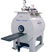
 7.0T Bruker BioSpec® USR (picture at left) magnet equipped with cryoprobe technology for high spatial resolution imaging of small models.
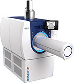
3T Bruker Biospec® (picture at right) featuring a motorized transport system; includes multiple RF coils.
3.0T Siemens Skyra wide-bore magnet available for imaging medium to large models.
Magnetic Resonance imaging (MRI) is an essential research tool for the non-invasive study of structure and function. The ability to use this technology in pre-clinical models allows translational studies that are crucial to elucidating a variety of disease mechanisms and to developing novel treatment approaches. From the study of brain function in neuropsychiatric disease, to the measurement of pulmonary function, to the introduction of new, minimally invasive image-guided treatment approaches, the use of novel MRI research methods is revolutionizing biomedical science.
 Optical Imaging
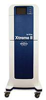
The Bruker In-Vivo Extreme II Optical/X-ray system provides both X-ray and full spectrum bioluminescence and fluorescence imaging capabilities. The system enables noninvasive and longitudinal monitoring of disease progression and response to disease-specific interventions. The ability to rapidly acquire both high-resolution and dynamic images make the Xtreme II an ideal system for multi-model pharmacodynamic studies and assessment of targeted clinical treatments, such as tracking tumor and metastases progression, in a variety of preclinical models. Key features include an ultra-sensitive 4MP CCD camera capable of high-resolution imaging (to 13.5 microns), 28 excitation filters ranging from 400nm to 780nm, and 6 wide-angle emission filters ranging from 518nm to 850nm.
 Micro-CT
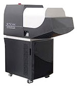
The Bruker Skyscan1176 provides for ultra-low X-ray dose in vivo scans for longitudinal studies with minimal radiation effects. In vivo imaging can be performed at nominal resolutions of 9, 18 or 35 microns with a maximum FOV of 68mm x 200mm. With the ability to alter beam energy, exposure time, and filtering this system is excellent for both soft tissue and bone imaging. One of the applications of the scanner is bone studies where the morphometric parameters such as bone mineral density, bone volume ratio and trabecular thickness can be extracted from the images. It is capable of monitoring bone architecture changes over time, evaluating possible healing treatments or regenerations and understanding skeletal development or deformity progress like rheumatoid arthritis.
Micro CT can also be used for various lung-disease studies including lung fibrosis. The low radiation dose as well as the ability to synchronize scans with respiratory cycles provides excellent in vivo longitudinal lung imaging.
 PET/CT
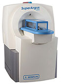
Sedecal SuperArgus PET/CT 4R: LYSO/GSO phoswich detector PET/CT; 8.3% sensitivity, spatial resolution < 1mm across entire FOV; transaxial FOV 120 mm, axial FOV 100/350 mm without/with translation; simultaneous imaging of up to 4 mice; integrated anesthesia, heated bed, gating, monitoring.
The SAIL staff collaborates with investigators who wish to use the imaging lab in planning the most appropriate and efficient use of the facility. For more information on the center and collaboration opportunities, contact the SAIL team at SAILinfo@partners.org.
Q. What is the price of performing scans?
A. Information on pricing can be found on the BWH Research Imaging Core website.
Q. Can I tour the facility?
A. Yes, we would love to show investigators and staff our facility. Please contact the SAIL Research Coordinator, at SAILinfo@partners.org to set up an appointment.
Q. Is it possible to obtain pilot data for grants so that I may receive funding in the future?
A. Researchers without a source of funding may collect pilot data to include in a grant. To apply for pilot data time please contact the SAIL Research Coordinator at SAILinfo@partners.org.
For over a century, a leader in patient care, medical education and research, with expertise in virtually every specialty of medicine and surgery.
About BWH

