Uterine fibroids are benign (not cancerous) tumors that arise from the muscle layer (myometrium) of the uterus. The uterus is mostly made out of smooth muscle cells, designed to expand with the growing pregnancy and to help with vaginal delivery by contracting forcefully at the end of pregnancy. Uterine fibroids can grow underneath the endometrium (submucosal), in the myometrium (intramural) or underneath the outer lining (serosa) of the uterus (subserosal).
Uterine fibroids are very common and are found in about 25% of women between the ages of 18-45. When examining surgically removed uteri from women of all ages, up to 80% of women are found to have some fibroids present. African American women have higher risk of developing uterine fibroids compared to white women. Other risk factors for developing uterine fibroids include nulliparity (never carried a pregnancy beyond 20 weeks), heavy alcohol drinking, and genetics. It has also been proposed that high consumption of red meat and ham could increase the risk of fibroids, while consumption of fruits and vegetables could decrease the risk of fibroids, however this has not been conclusively proven. Consistent use of birth control pills seems to lower the risk of developing uterine fibroids.
It is not well understood why fibroids develop. There are some genetic abnormalities in the smooth muscle cells that increase the risk and the fibroids seem to be under hormonal control, although the precise mechanism of this is not fully understood either.
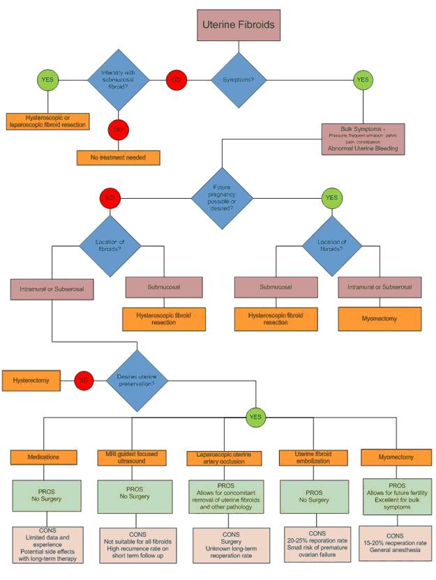
Uterine fibroids can cause abnormal uterine bleeding, pelvic pressure and can have a negative effect on fertility. In general, fibroids that press on the endometrium can cause abnormal bleeding. This includes submucosal fibroids and large intramural fibroids. When the fibroids grow larger the uterus expands and puts pressure on organs that are close by, such as the urinary bladder and the rectum. This can lead to a feeling of fullness, constipation and frequent urination. In extreme cases the uterus can be the size of a full term baby, reaching all the way up to the liver and diaphragm. Fibroids usually do not cause extreme pain, however sometimes a fibroid might lose it's blood supply due to rapid growth. This causes the fibroid to die (degenerate) which can be very painful. The pain will usually get better over a period of few days, but sometimes requires a surgical intervention.
Fibroids that press on the endometrium probably decrease the chance of getting pregnant since this can interfere with the implantation of the embryo. Unfortunately, there are no carefully designed scientific studies that evaluate the effects of fibroids on fertility, however based on the best available evidence, fibroids that distort the uterine cavity should be removed in patients with infertility problems.
Large fibroids can usually be identified during a regular physical exam. Ultrasound can identify most fibroids and is easily performed in a doctors office. The addition of saline infusion sonohysterography can help to identify fibroids that are inside the uterine cavity. Office hysteroscopy is also an excellent tool to detect uterine fibroids inside the uterine cavity.
Magnetic resonance imaging (MRI) gives the best information about the exact location and size of the fibroids. It is a very useful tool to determine appropriate treatment options, such as removal of the uterine fibroids (myomectomy), embolization or focused ultrasound (see below). MRI can also be helpful in determining if there is something else in the uterus that is causing the symptoms, such as adenomyosis or a leiomyosarcoma (cancer of the uterine muscle). It is however the most expensive diagnostic option and should be used selectively.
It is important to note that since fibroids are not cancerous, no treatment at all is probably the best option for women who have no symptoms associated with the fibroids. The traditional treatment for uterine fibroids is a large abdominal incision and either removal of the uterus (hysterectomy) or removal of the fibroids (myomectomy).
Hysterectomy has usually been recommended for women who are not planning to have any more children, since there is a 15-20% risk that the symptoms might not improve following a myomectomy, which will require additional surgery. However, there are several women who want to conserve their uterus even though they are not going to have any more children. It is important to respect their wishes as long as they fully understand the risks and benefits associated with their decision. The majority of women with uterine fibroids will not need to have a laparotomy to fix their problem. We will discuss some of the minimally invasive and non-invasive treatment options that are currently available.
The growth of uterine fibroids is controlled by estrogen and progesterone, two of the more common female hormones. By turning off the production of these hormones or reducing the influence they have on the fibroids, it is possible to reduce the size of the fibroids significantly. Gonadotrophin releasing hormone (GnRH) is normally produced in the brain and controls the production of estrogen and progesterone. The secretion of this hormone varies from time to time, i.e. it is cyclical.
When a synthetic version of GnRH (called GnRH agonist) is given to women, there is an initial 2 week period of stimulation of hormone production, but then the system becomes oversaturated and the production of estrogen and progesterone shuts down temporarily. GnRH agonists are usually injected into muscle in long-acting formulations that have an effect for one or three months. They can also be given using a daily nasal spray. Common GnRH agonists include Lupron, Zoladex and Synarel. In three months, GnRH agonists can shrink to uterine size by one third and the fibroid size by half. In addition, vaginal bleeding usually stops completely.
Symptom relief from pressure or bleeding is therefore common. Unfortunately, GnRH agonists have significant side effects, such as nights sweats, hot flushes, irritability, vaginal dryness and difficulty sleeping. In addition, long term use is associated with significant bone loss (estrogen stimulates bone formation). Because of this it is not recommended to use GnRH agonist longer than 6 months. It is possible to reduce the side effects of the GnRH therapy by using a very low dose of estrogen or progesterone (add back therapy).
It is generally recommended to wait at least four weeks before starting the add back therapy, in order to get a maximum effects on the fibroids. Because GnRH agonist therapy can only be used for a short period of time and because the effects are quickly reversed, GnRH agonist therapy is mostly used to reduce the size of uterine fibroids before surgery. This will make it easier for the surgeon to remove the uterus or fibroids at the time of surgery. The maximal effect on uterine size is reached at three months and therefore it is not necessary to treat women longer than that preoperatively.
There is an intense search going on for a suitable long term medical treatment for fibroids. One promising option seems to be medications call selective progesterone receptor modulators (SPRM). These medications interact with cell receptors that respond to progesterone and change the effect that progesterone has on cells. One of these medications called Mifepristone has been found to be effective in reducing the size and symptoms of fibroids. However, it is associated with side effects such as endometrial hyperplasia and abnormal liver enzymes. Another SPRM that shows promise is Asoprisnil. Early studies indicate that this is effective with no effect on the endometrium or liver. Time will tell if these and other medications will be effective, but the early results are very promising.
Birth control pills do not seem to be very helpful to treat symptoms associated with uterine fibroids, however they are effective in treating abnormal bleeding due to problems with ovulation. It is possible that the abnormal bleeding that the woman is experiencing might not be due to the fibroids and therefore a 3 month trial of birth control pills is appropriate in select cases.
The hormonal IUD might be effective where the uterine fibroids are small, however in the case of large fibroids it is probably not effective for symptomatic control.
Myomectomy should be the surgical option of choice in women who want to have more children. Other options such uterine artery embolization, uterine artery occlusion, cryomyolysis and focused ultrasound are usually not recommended for women who want to have more children. There are nevertheless some women who have become pregnant after these procedures and successfully carried their babies to term without complication. Most of these women had uterine artery embolization.
The number of complications associated with the pregnancies that occurred in these women was significantly higher than for women who did not have these procedures. Some of that might be explained by the fact that the women who underwent these procedures had other health problems that might make it more likely for them to have problems during their pregnancy. Nevertheless, it seems clear that the risk of complications is increased and that women who want to become pregnant should have either an abdominal or a laparoscopic myomectomy.
Myomectomy or removal of fibroids is commonly performed in women who want to retain their fertility. It is possible to remove most fibroids laparoscopically, however there are certain limitations such as size and location that we will discuss. It depends largely on the surgeon's skill and experience what cases can be performed laparoscopically. In general fibroids that are larger than 10cm and are inside the uterine muscle (intramural) can be difficult to remove. Also, if there are many fibroids (>5) it can be difficult to complete the surgery laparoscopically.
Fibroids that are located next to large blood vessels or in the cervix can also be challenging to remove laparoscopically. A main limiting factor is laparoscopic suturing. Suturing laparoscopically is a very advanced skill and most gynecologists do not have the capability to do this properly. It is critical to close the uterine incision adequately, because otherwise the risk of uterine rupture during pregnancy is increased. Remember that most women who have a myomectomy are hoping to carry a pregnancy to term after the procedure. If the scar on the uterus is weak, it can rupture when the pregnant uterus has become very large and is contracting during labor. This can be very dangerous for the mother and the baby.
A myomectomy is usually performed in the following manner. An incision is made into the uterine serosa (outer lining of the uterus) and carried all the way into the fibroid. The fibroid is then gently freed from the uterus by pulling on it and cutting away it's attachments. Once the fibroid is removed the uterine incision needs to be closed. This portion is the most challenging part when performing the procedure laparoscopically, because it involves a lot of suturing. The uterine incision is closed in layers, usually 3-4 depending on the depth of the uterine incision. The fibroid itself is removed via the abdominal incision.
Because suturing is the most challenging part during a laparoscopic myomectomy some modifications have been developed. One is called a laparoscopically assisted myomectomy (LAM). During this technique the fibroid is released laparoscopically as previously described and then a small laparotomy incision (5-6cm) is made above the pubic hairline. The fibroid is removed through this incision and the uterus is sutured through it as well. This technique is well suited when dealing with a large number of fibroids or when physicians are not proficient in laparoscopic suturing. It is not well suited for fibroids that are located on the back of the uterus, since it is difficult to reach this area through a small incision.
Another alternative is using the da Vinci robot for the myomectomy. The da Vinci robot consists of 3 to 4 mechanical arms that are controlled by the surgeon from a separate control unit (console). The arms of the robot can carry various instruments and the instruments can move freely at the tip, allowing much more freedom of movement than with traditional laparoscopy. In addition, the surgeon has three dimensional vision of his environment through the control unit. This makes suturing much more easier and allows more surgeons to be able to complete the case laparoscopically.
The robot has some disadvantages as well, including lack of tactile sensation, i.e. the doctor is not able to feel how hard or soft an organ is or how tightly a knot is being tied and has to rely completely on what he sees. The robot is also very bulky and adds about 30 minutes to the duration of the surgery because of the amount of time it takes to set it up and remove it following the case. It is also very expensive and the current version uses slightly larger trocars than the trocars used in standard laparoscopy cases, which makes the scars a little bigger and perhaps increases post-operative pain.
In appropriately selected patients, uterine artery embolization (UAE) can be an effective treatment option, especially for women who for a variety of reasons are not good candidates for surgery. Uterine artery embolization is performed by an interventional radiologist. A small catheter is placed into a blood vessel in the groin and it threaded up to the uterine artery under x-ray guidance. Once the catheter is in the right place, small particles are released into the blood stream that subsequently become stuck in the blood vessels supplying the fibroids. This reduces or stops blood flow to the fibroids, which in turn causes them to die and shrink. This technique is also sometimes called uterine fibroid embolization (UFE), since it has become more selective and targets individual fibroids rather than the whole blood supply of the uterus.
The UFE procedure itself usually takes about one hour to complete. UFE is very effective in properly selected patients, with over 85% of patients reporting significant improvement in symptoms, even up to 5 years after treatment. Patients with multiple fibroids or fibroids larger than 8cm usually don't do quite as well. In addition, patients with submucosal fibroids are not good candidates for this procedure, since treatment failure is high and there is some risk of developing a serious infection. Patients with very large fibroids and a lot of pressure symptoms usually do not get completely better from their pressure symptoms after UFE. Finally, patients who have a large subserosal fibroid on a narrow stalk (pedunculated fibroid) are not good candidates for UFE.
Following the procedure there is commonly a lot of pain and discomfort. This is due to the necrosis (dying) of the fibroids, which causes inflammation and swelling of the uterus. Patients are usually admitted for one or two days following UFE for pain control. Fever is common (due to the inflammation) and some patients experience nausea. These symptoms gradually improve over time and most patients are back to normal in about two weeks.
When compared with abdominal myomectomy the two treatments were equally effective, but patients who had UFE had a shorter hospital stay (one versus 2.5 days) and were able to return to normal activities quicker (15 vs 44 days). Another comparative study however found that more patients in the UFE group needed additional surgery and did not have as good relief of their symptoms. To date there have been no properly designed studies that compare UFE to laparoscopic myomectomy.
This is an alternative to UFE, where the uterine artery is located and permanently clamped using laparoscopy. The uterus regains it's blood supply within 6 hours, however the fibroids are not able to do this and die off. A recent comparative study between UFE and LUAO found the two procedures to be equally effective in reducing bleeding when measured with pictorial charts. However, more women in the LUAO group complained of excessive bleeding six months after the procedure than in the UFE group. There was significantly less pain associated with the LUAO than UFE. LUAO is a promising alternative to UFE, especially when there are large fibroids or pedunculated fibroids present. This allows the physician to remove the large fibroids during the LUAO procedure, which might help better to relieve pressure symptoms than uterine fibroid embolization.
This is an experimental technique where the uterine arteries are located vaginally with the help of ultrasound and temporarily clamped for six to eight hours. As discussed before, when the uterine artery is clamped, the uterus regains it's blood supply within 6 hours, however the fibroids are not able to do this and die off. The patients are usually offered an epidural for pain relief and are temporarily admitted to the hospital while the clamp is on the uterine arteries. Once the clamp is removed patients are able to go home the same day. The preliminary results are promising, however more studies are needed before this becomes available as a treatment option to the general public.
This technique involves localizing the uterine fibroids laparoscopically and destroying them with either extreme heat or cold. Usually a needle is inserted into the fibroid and the tip of the needle is heated or cooled to destroy the fibroid. Preliminary studies indicate significant reduction in fibroid volume and also significant improvement in symptoms. The ideal candidate should have no more than 4 fibroids and no fibroid larger than 10 cm. The advantage over myomectomy is that here no suturing is required. However the destruction of the fibroid can result in the formation of a lot of scar tissue and possible a weak uterine wall. Myolysis is therefore not recommended for women planning to have more children and is considered experimental at present.
This is an outpatient procedure, where an MRI is used to locate the fibroids and multiple ultrasound beams are focused on a small portion of the fibroid at a time. The focused ultrasound waves create a lot of heat which destroys the uterine fibroid. The procedure takes place in a radiology suite and the patient lies prone on an MRI table. The procedure time is two to three hours. Early studies are promising for this technique, however it is not a good option for women who want to have more children.
Other patients who are not well suited for this procedure are patients with submucous fibroids, multiple fibroids or fibroids near the bowel or bladder. Magnetic resonance guided focused ultrasound is also a very expensive procedure and should be used only on selected patients initially until better long term follow-up and results are available.
To schedule an appointment, call (617) 525-8582 or visit our contact information page.
Read Dr Einarsson's publication on laparoscopic myomectomy for management of symptomatic fibroids.
For over a century, a leader in patient care, medical education and research, with expertise in virtually every specialty of medicine and surgery.
About BWH