Renal imaging provides your doctor with functional images of your kidneys. That means the images show how the cells are performing—whether normally or abnormally, and to what degree. Blood flow can also be examined. There are several kinds of studies, but the processes are similar. This is how they work:
Renal imaging can be used for a variety of reasons. Diuretic renal scintigraphy is used to detect kidney blockage. Cortical renal scintigraphy is used to measure the amount of functioning kidney tissue you have. Renal perfusion imaging with pharmacologic (captopril) intervention is also used to evaluate the blood flow to the kidneys and to detect probable narrowing of the renal arteries. Your doctor will determine which renal imaging procedure is necessary.
Your doctor will schedule the examination. When the exam is scheduled, your doctor must send a written order detailing the type of exam you should receive. You should also request a copy of this written order, and you should bring it with you to your appointment. According to Massachusetts state law, the technologist cannot inject the radiotracer without a written order from your doctor. Your examination will be delayed if we do not receive a written order.
When your examination is scheduled, your doctor will be told the time for your injection, the time for imaging, and the expected length of time for the complete examination. These times are provided to help you plan your visit to our laboratory.
As you plan your visit to our lab, please remember that children under the age of 12 are not permitted within the Nuclear Medicine waiting room or laboratory areas.
The preparation will depend on the type of exam, and you will receive specific instructions from your doctor. For some studies, a diuretic – a drug that causes increased urine production by releasing water from body cells – may be administered during the exam. For some, a catheter may be inserted to keep the bladder empty. In some cases, you may be asked to discontinue prescribed medications. You will be asked to drink plenty of water before your exam, and to void before the test begins.
The radiotracers are not known to cause any side effects or adverse interactions with food or prescription drugs.
Prior to the administration of the radiotracer, any woman between the ages of 12 and 56 will be asked if she might be pregnant. If you think you might be pregnant, you should talk to your doctor about it before having a nuclear medicine examination.
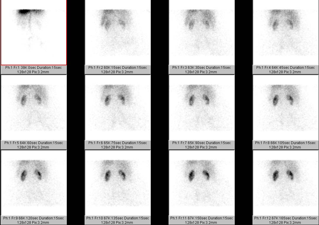
During the actual imaging portion of any exam, the technologist will ask you to lie on your back on a bed positioned between a set of cameras. Once you are comfortable on the bed, the imaging will begin. It is important that you remain still, because movement can ruin the images, and the nuclear medicine physician may have difficulty interpreting them accurately.
After the scan is complete, you will be able to resume normal daily activities. There will be no restrictions on eating or drinking. The radiotracers do not cause drowsiness, so you will be able to drive.
A nuclear medicine physician will review and interpret the images obtained during your study. The results of your renal imaging study will be provided to your doctor within 48 hours.
Most people do not experience any side effects from the radiotracer. Hypotension (drop in blood pressure) might occur in those patients taking captopril medication before the renal scan for renal artery stenosis begins. However, this drop in blood pressure responds to the intravenous administration of normal saline. Your blood pressure will be monitored every 15 minutes for 1 hour.
Nuclear medicine examinations do involve the use of a small amount of radiation. The tracer dose is calculated to minimize radiation exposure while providing accurate test results.
Nuclear medicine studies may not be appropriate for pregnant women or those who are breastfeeding. If you think you may be pregnant, discuss this with your doctor. Of course, it is always important to consider the benefits of any diagnostic study along with the risks. In some cases, the importance of making the correct diagnosis outweighs the potential risk to the unborn baby. Your doctor can explain your options.
If you are breastfeeding, you should not nurse your baby for approximately 36 hours after the radiotracer injection, since radiation can be passed through the breast milk. Prepare for your examination by pumping and saving milk for 24-48 hours before your examination, then bottle-feed your baby during the hours following your appointment.
The radiation administered during a nuclear medicine study is eliminated from you body through the kidneys. For that reason, you should drink plenty of fluids and urinate frequently following your examination.
Glossary terms
For over a century, a leader in patient care, medical education and research, with expertise in virtually every specialty of medicine and surgery.
About BWH
