Staff
|
Michael Steigner, MD
Director
|
Dimitris Mitsouras, PhD
Director, MR Physics and Engineering
|
|
Kanako Kumamaru, MD, PhD
Assistant Professor
|
Amir Imanzadeh, MD
Research Fellow
|
|
Tianrun Cai, MD
Research Fellow
|
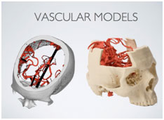
Vascular imaging using ultra-high resolution three-dimensional MRI of the vasculature
Our approach, termed “inner-volume reduced field-of-view non-selectively refocused 3D fast spin-echo” is based on surpassing a main limitation of MR imaging systems by enabling them to concentrate imaging time on only the vasculature being assessed, rather than sharing imaging time with other tissues that are not of interest.
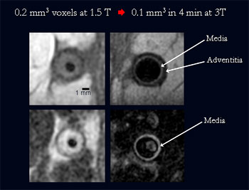
Inner-volume 3D MRI enables an MR system to “zoom in” to the vessel of interest. The improved spatial resolution in clinical patients gives new opportunities to examine physiologic states such as outward remodeling or measurements of neo-intimal thickness and hypertrophy of the media. We have focused our research efforts in patients with peripheral vein bypass grafts. |
Clinical Applications
We have an ongoing interest in the following:
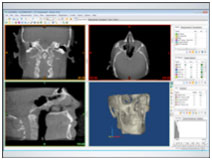
- Image post-processing in cardiovascular imaging
- Optimizing the radiation dose for patients who require
- CT as part of their clinical evaluation.
- Computed tomography for pulmonary embolism
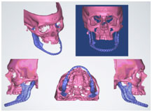
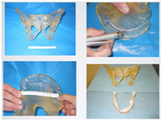
3D Printing
The AISL is a leader in developing tools that can be used for 3D visualization in traditional 2D formats, as well as 3D printing of graspable objects, all from the same segmentation. This work will streamline diagnoses and surgical planning in patients for whom the 3D printed model is helpful for planning an intervention. We have studied these methods for face transplantation.
Training
Providing research opportunities for enthusiastic trainees is a trademark of the AISL. Many radiology and cardiology residents and fellows who have come to the lab have gone on to productive academic careers. We are interested in teaching sound methods basic science and clinical research.
Downloadable Materials