Brigham and Women's Hospital has a rich history of pioneering technologies and imaging tools to deliver the most advanced care to patients.
Today, clinicians at the Brigham are applying the latest innovations in imaging to support world-class research and improve the care of patients across a variety of disciplines.
"As a major academic medical center with significant funding from industry and government, we are translating scientific discoveries into clinical care as fast as we can," said Giles W. Boland, MD, chair of the Department of Radiology. "In many cases, the Brigham invented these technologies, and now we're translating them into patient care—right at the forefront of where much of medicine is going."
In September 2018, a 7 Tesla (7T) MRI scanner became available for patient care at the Brigham. The advanced diagnostic imaging tool has more than double the strength of a conventional high-field scanner.
The Brigham's 7T is part of a new generation of ultra-high field instruments and only the second to be approved for clinical use in the United States. When it arrived in May 2017, the 25-ton system was lowered by crane into a space built to accommodate it.
"The 7T MRI gives us a much higher detection rate than the conventional 3T and 1.5T; it's a much more powerful magnet that gives us extremely high-resolution images," Dr. Boland said. "This is going to transform the investigation and diagnosis of neuroinflammatory diseases, particularly MS and Alzheimer's, as well as epilepsy. It also has applications for musculoskeletal joint imaging."
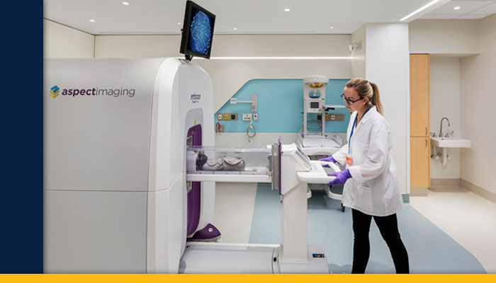
A new MRI system arrived in the Department of Pediatric Newborn Medicine in September 2018. Specifically designed for the safe imaging of newborns, it's the world's first FDA-approved, NICU-dedicated MRI system.
Babies undergoing scans are placed in a temperature-controlled, self-contained incubator bed that minimizes their movement while allowing for better control of their environment and continuous monitoring of their vital signs. Information gained from the MRI can inform clinicians and the family as to whether brain injury has occurred, and in the future, guide which treatments may assist in preventing disability.
"Having this MRI in the NICU will reduce time and patient risk associated with transporting newborns to a traditional MRI and allow MRI access from the first hours of life through the challenging, sometimes life-threatening, time within the NICU," said department chair Terrie Eleanor Inder, MBCHB.
The hybrid operating room at the Heart & Vascular Center now features the ARTIS Pheno angiography system. The state-of-art system is used for patients who require hybrid cardiovascular interventions combining intravascular and open surgical procedures.
"This next-generation angiography system allows us to do these complex hybrid procedures more safely and efficiently. Both patients and doctors are also exposed to less radiation compared with earlier systems," said Michael Belkin, MD, chief of the Division of Vascular and Endovascular Surgery. Furthermore, the Pheno system provides resolution that is four times higher for two-dimensional imaging than previous iterations.
Early work in MRI-guided focused ultrasound (MRgFUS) thalamotomy took place at the Brigham 20 years ago. Now this noninvasive technology is being used to treat patients with medically refractive essential tremor.
"We very accurately and precisely ablate the specific nucleus in the thalamus to dramatically reduce tremor," said G. Rees Cosgrove, MD, FRCSC, a stereotactic and functional neurosurgeon and director of epilepsy and functional neurosurgery at the Brigham, one of only 11 MRgFUS treatment centers in the United States and the only one in New England.
Meanwhile, Alexandra J. Golby, MD, director of image-guided neurosurgery, is using cutting-edge imaging techniques to assist with laser interstitial thermal therapy (LITT). This therapy involves inserting a laser into a tiny hole in the skull, which makes it much less invasive than a craniotomy.
With LITT, the laser heats up and destroys tissue such as brain tumors, areas of necrosis or areas causing seizures. Continuous imaging is performed during the therapy to measure the temperature change as the laser is being applied. Following the procedure, final imaging is done to document the results.
Moving forward, the Department of Radiology will continue to leverage the latest advancements in imaging to enhance patient care, including in our world-renowned Advanced Multimodality Image Guided Operating (AMIGO) Suite. Another point of focus is using artificial intelligence to read scans more effectively and integrate the massive amounts of data residing in EMRs, thus helping doctors make more-informed decisions with better patient outcomes.
"We're at the forefront of exploring innovative ways to transform health care so that we can get the right treatment to the right patient at the right time," Dr. Boland said. "Artificial intelligence is a key part of the strategy to help us get there."

