Overall Goals of the Program
The Quantitative Musculoskeletal Imaging Group (Q-MIG) in the Radiology Department at Brigham and Women’s Hospital specializes in developing software image analysis methods to quantify structural changes related to musculoskeletal diseases. The primary focus is on osteoarthritis (OA) but we have substantial experience with rheumatoid arthritis (RA) and with various animal models to investigate more general disease processes. The overall goal of the program is to provide clinical researchers with fast and objective methods that can facilitate high-powered studies and clinical trials.
Program History
The program was initiated by Dr. Jeffrey Duryea in 2002, building on the research efforts that began when he was on the faculty of at the University of California, San Francisco. The early work, supported by National Institutes of Health (NIH), Whitaker Foundation, and Arthritis Foundation grants, focused on methods to characterize hand RA radiographically. Later, software tools were developed to study knee OA for both radiographic images and MRI data sets.
The Q-MIG group is a formal collaborator of the Osteoarthritis Initiative (OAI), a large NIH and industry funded study of OA that has collected a substantial amount of images and clinical data from OA subjects. The data from this study have formed the backbone of the more recent research efforts by the group supporting the majority of grant applications and publications. Additionally the group was selected by the OAI to perform software readings of a large number of knee radiographs (over 35,000 to date) that are released to the OA research community and have supported many research projects outside of Brigham and Women’s Hospital.
Accomplishments
The majority of the work during the past five years has been supported by a successful NIH grant. (R01AR056664). The goal of this study was to develop and validate several software-based measures of knee OA from MRI data sets. Much of the group’s recent effort has been directed at accomplishing the aims of this grant proposal. We now have fast quantitative methods in place for cartilage morphometry (1-2), bone marrow lesion (BML) (3), and osteophyte volume (4), and have looked at associations of these measures with pain. Methods are in place to assess large numbers (several hundred) subjects for multiple disease-related structures, which will facilitate future higher power studies of knee OA. We are actively seeking additional funding to support the next phase of the program.
Additionally we are pursuing several different but related research projects. Methods have been developed and validated to characterize progression the progression of hip and hand OA radiographically (5). In collaboration with Dr. Chris Evans at the Mayo Clinic, Dr. Duryea was recently awarded a supplemental grant to support a study testing the hypothesis that a novel method based on conventional radiography can be used to detect the status of osseous defects with high accuracy. We have also received a NIH grant to support a study to evaluate hand radiography in conjunction with a study of adiposity-related metabolic factors as predictors of incident hand OA. In collaboration with Daniel Solomon in the BWH Rheumatology Division, Department of Medicine we are developing methods to assess hand RA using multi-slice CT. With Drs. Anthony Aliprantis and Matthew Harris, we have initiated a project to apply quantitative methods to assess bone parameters of zebra fish.
References
- Iranpour-Boroujeni T, Watanabe A, Bashtar R, Yoshioka H, Duryea J. Quantification of cartilage loss in local regions of knee joints using semi-automated segmentation software: analysis of longitudinal data from the Osteoarthritis Initiative (OAI). Osteoarthritis Cartilage. 2011;19(3):309-14. PMCID: 3046247.
- Duryea J, Iranpour-Boroujeni T, Collins JE, Vanwynngaarden C, Guermazi A, Katz JN, Losina E, Russell R, Ratzlaff C. Local-area cartilage segmentation (LACS), A semi-automated novel method of measuring cartilage loss in knee osteoarthritis. Arthritis Care Res (Hoboken). 2014.
- Ratzlaff C, Russell RL, Duryea J. Quantitatively-measured bone marrow lesions in the patellofemoral joint: distribution and association with pain. Osteoarthritis and Cartilage (abstract). 2014;22, Supplement:S247-S8.
- Hakky M, Ratzlaff C, Guermazi A, Duryea J. Responsiveness of a semi-automated method to measure osteophyte volume change in knee osteoarthritis over four years using 3.0 T DESS 3D MRI. Osteoarthritis and Cartilage (abstract). 2013;21, Supplement:S202-S3.
- Ratzlaff C, Van Wyngaarden C, Duryea J. Location-specific hip joint space width for progression of hip osteoarthritis: Data from the osteoarthritis initiative. Osteoarthritis Cartilage. 2014;(in press).
Personnel and Collaborators
Current Q-MIG Personnel
- Jeffrey Duryea, PhD - Director
- Quinley Miao, BA
- Lena Schaefer, MD
- Stacy Smith, MD - Clinical Co-director
- Meera Sury, BA
- Ming Yin, BA, MS
Radiology Department Collaborators
- Bruno Madore, PhD
- Jeremy Wortman, MD
- Violeta Nikac, MD
Brigham and Women's Hospital Collaborators (outside of the Radiology Department)
- Jeffrey Katz, MD (Orthopedics)
- Julia Charles, MD (Medicine, Rheumatology)
- Daniel Solomon, MD (Medicine, Rheumatology)
Collaborators Outside Brigham and Women's Hospital
- Chrisopher Evans, PhD (Mayo Clinic)
- Timothy McAlindon, MD (Tufts University Medical School)
- Jeffrey Driban, PhD (Tufts University Medical School)
- Charles Eaton, MD (Brown Universty)
- Kent Kwoh, MD (University of Arizona)
- Michael Nevitt, PhD (U.C. San Francisco)
- Tuhina Neogi, MD Ali Guermazi, MD (Boston University Medical School)
- Matthew Harris, PhD (Children's Hospital)
- Martin Englund, MD PhD (Lund University, Sweeden)
- Joanne Jordan, MD, Amanda Nelson, MD (University of North Carolina)
Funded Projects
- R01AR056664, "Quantitative MRI analysis method for longitudinal assessment of knee OA"
- R01AR050243, "Healing of segmental defects of bone by gene transfer"
- R01AR066378, "Constitutional and metabolic factors associated with the development of Hand OA"
Radiographic JSW at fixed locations
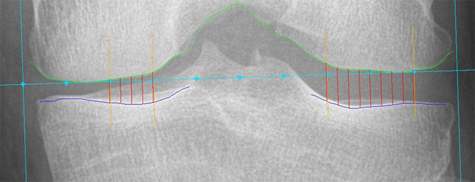 |
Hand JSW Measurement
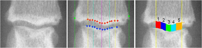
Five Measurement regions: JSW1, JSW2, JSW3, JSW4, JSW5 Central JSW measurement: JSWC = (JSW2+JSW3+JSW4)/3 |
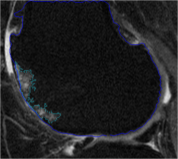
Bone marrow lesion volume
 |
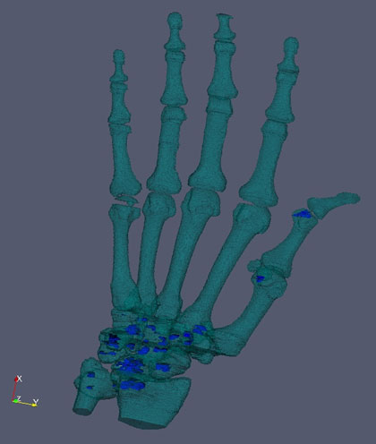
Hand CT to evaluate RA erosions
 |
Zebrafish vertebrae morphometry
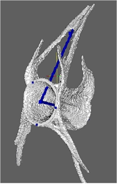 |


