PET stands for positron emission tomography. It is an imaging technology used in nuclear medicine to obtain pictures of the metabolic (functional) activity within particular areas of the body. The concept of a PET scan is similar to other types of nuclear medicine examinations:
Because PET scanning "sees" chemical activity in various tissues, it can reveal many disorders much earlier than other imaging techniques. PET scanning may be used in conjunction with other imaging techniques to provide a direct correlation between anatomy and function.
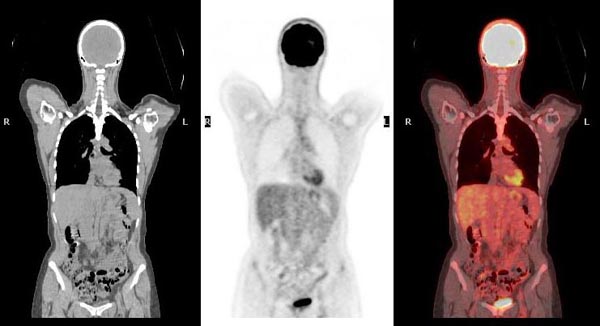
PET scanning is used to detect cancer, as well as a number of cardiovascular and neurological abnormalities.
Cancer cells, for example, have a much higher level of metabolic activity than normal cells. PET scanning detects that increased metabolic rate and can therefore:
PET is considered to be particularly useful in evaluating lung, head and neck, colorectal, esophageal, lymphoma, melanoma, breast, thyroid, cervical, pancreatic, and brain cancers.
Certain neurological disorders have characteristic metabolic changes. These are readily apparent on PET images. Alzheimer’s disease, for example, has a unique appearance on PET scans. This appearance can differentiate Alzheimer’s from other forms of dementia, such as Pick’s disease, and give the patient earlier access to appropriate treatments.
In cardiovascular disease, PET images provide information about blood flow and the presence of damaged muscle tissue. This information is key to planning appropriate treatments and disease management.
Your doctor will schedule the examination. When the exam is scheduled, your doctor must send a written order detailing the type of exam you should receive and the region of interest. You should also request a copy of this written order, and you should bring it with you to your appointment. According to Massachusetts state law, the technologist cannot inject the radiotracer without a written order from your doctor. Your examination will be delayed if we do not receive a written order.
When your examination is scheduled, your doctor will be told the scheduled times of particular parts of the examination, such as the time of injection, the time of imaging, and the expected length of time for the complete examination. These times are provided to help you plan your visit to our laboratory. It is possible that your imaging appointment could change by as much as 1 hour, based on activity in the lab on the day of your appointment.
As you plan your visit to our lab, please remember that children under the age of 12 are not permitted within the Nuclear Medicine waiting room or laboratory areas.
Your preparation will depend on the purpose of the scan as described by your physician. You will receive instructions when your appointment is scheduled.
Prior to receiving her injection, any woman between the ages of 12 and 56 will be asked if she might be pregnant. If you think you might be pregnant , you should talk to your doctor about it before having a nuclear medicine examination.
A technologist will inject an appropriate dose of radiotracer. Depending on the purpose of the scan, you may be asked to wait before images can be taken. When it is time for the imaging session, the technologist will position you on a "bed" (also called a "couch") that is attached to the PET scanner. The scanner itself looks like a large box with a central, round opening. Gamma ray detectors are located all around the opening. The bed will slide into the opening, and the detectors will record the gamma rays being released from the body from your head to your feet. If you are severely claustrophobic, you may ask your doctor for an appropriate prescription to help you relax.
The imaging portion of the examination typically lasts 30 minutes to 1 hour. It is important that you remain still while the cameras are on—movement can ruin the images, and the radiologist may have difficulty interpreting them accurately.
After the scan is complete, you will be able to resume normal daily activities. There will be no restrictions on eating or drinking. The radiotracers do not cause drowsiness, so you will be able to drive.
A nuclear medicine physician will review and interpret the images obtained during your study. The results of your PET scan will be provided to your doctor within 48 hours.
Most people do not experience any side effects from the radiotracer.
Nuclear medicine examinations do involve the use of a small amount of radiation. The tracer dose is calculated to minimize radiation exposure while providing accurate test results.
Nuclear medicine studies may not be appropriate for pregnant women or those who are breastfeeding. If you think you may be pregnant, discuss this with your doctor. Of course, it is always important to consider the benefits of any diagnostic study along with the risks. In some cases, the importance of making the correct diagnosis outweighs the potential risk to the unborn baby. Your doctor can explain your options.
If you are breastfeeding, you should not nurse your baby for approximately 36 hours after the radiotracer injection, since radiation can be passed through the breast milk. Prepare for your examination by pumping and saving milk for 24-48 hours before your examination, then bottle-feed your baby during the hours following your appointment.
The radiation injected during a nuclear medicine study is eliminated from you body through the kidneys. For that reason, you should drink plenty of fluids and urinate frequently following your examination.
Glossary terms
For over a century, a leader in patient care, medical education and research, with expertise in virtually every specialty of medicine and surgery.
About BWH
